Applications of Sub-nanosecond Lasers in Two-photon Fluorescence Microscope
Project Background
In the late 1980s, as the advanced biological fluorescence imaging technique in the world, laser confocal microscope has been widely applied in the field of fluorescence microscope imaging for biological materials. However, laser confocal microscope has a disadvantage,confocal aperture does not only block fluorescence outside of focal spot, but also the scattering fluorescence of biological tissue produced by focal spot. It leads to the decreased fluorescence collection efficiency, and the imaging depth could not be more than hundreds of micrometers.Thus, Two-photon fluorescence microscope technology appears in 1990s, which combines laser confocal microscope and two-photon stimulating technologies.The stimulating wavelength in the two-photon absorbing process is always set in the range of biological window at 680-1080nm wavelength, which avoids the damage of UV light to cell or living body and deeper penetration.
At present femtosecond Ti sapphire laser is widely used in two-photon fluorescence microscope,but the large size and high price which limit the development of two-photon fluorescence microscope imaging technology in the life sciences, chemistry, medical applications. Therefore, the sub-nanosecond solid-state laser with compact design and favorable price is applied for standard assembled laser source in the applications like medical diagnosis.
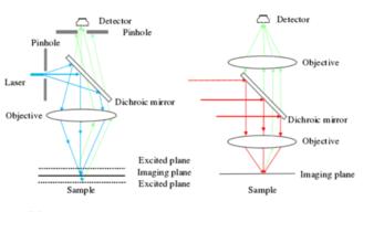 Difference between confocal microscope imaging/two-photon fluorescence imaging structure
Introduction to theory
The basic theory of two-photon excitation: In the high photon density, when fluorescence molecular is stimulated, the photons absorbed two long wavelength, and then return to ground state from spontaneous emission fluorescence photon after excitation transition and vibration relaxation.Comparing with traditional single-photon excitation fluorescence microscope imaging, the optical signal of two-photon excitation fluorescence microscope imaging is non-linear. The stimulated light is the laser source with long wavelength and high peak power, and the emission wavelength of fluorescence photon is a little longer than one half of the excitation wavelength.
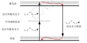
Two-photon excitation process diagram Two-photon fluorescence microscope imaging theory diagram
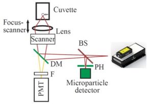
Technology and features
Two-photon fluorescence microscope imaging technique uses near-infrared laser source, and confocal aperture is not required due to its characteristics of non-linear. Comparing with confocal microscopy, thus two-photon fluorescence microscope imaging technique have the advantages as following:
Deeper imaging depth and less optical damage;
Confocal aperture is not required, and fluorescence collection efficiency is highly improved;
High spatial resolution and contrast ratio;
Small excitation range,phototoxicity and albifaction;
There is a long distance between emission wavelength and excitation wavelength, which avoids the spectral overlap;
Products and services
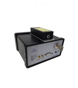
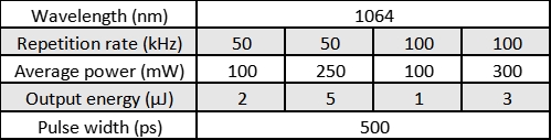
In the fields of two-photon fluorescence microscope, Reallight is available to offer different types of sub-nanosecond microchip laser with small size, single longitudinal mode and high peak power, to meet with the different customer’s requirements in the life sciences, analytical chemistry, clinical medicine applications. Meanwhile, RealLight can provide custom laser sources tailored to customers.
Disclaimer: Some content in this article is sourced from the internet for technical research and exchange purposes, and is provided for reference and learning. If there are inaccuracies or academic discrepancies, corrections are welcome. If there are copyright issues, please contact us for prompt verification and removal.